What is contrast sensitivity?
Contrast sensitivity refers to the ability to distinguish an object from other objects and the background, even when it is not clearly outlined or prominent. It is one of the ways that visual function is measured. Other ways of testing visual function include visual acuity, visual field and color vision.
Let’s use an example. Imagine a white-colored dog at a snow-covered park on a gloomy and dark winter evening. It wouldn’t be surprising if you had some trouble trying to find the dog regardless of how good your visual acuity is. Now imagine a black-colored dog at the same snow-covered park on a perfect sunny winter day. In this situation, you should have no problem spotting the dog.

Left: White dog in a white background (bottom left corner). The contrast here is low.
Right: Black dog in a white background. The contrast is high in this scene.
Maximum contrast is obtained when comparing between black and white. However, when comparing between shades of gray, your ability to discern outlines becomes reduced. Your sensitivity to contrast determines how well you can detect and determine outlines in lower contrast situations.
Why is contrast sensitivity important?
When you have poor ability to distinguish contrast, you will be unable to see small contrast differences between adjacent objects and surfaces. For example, you may find it difficult to see facial expressions. You may find navigating steps difficult because you are unable to recognize the edges of the steps.
Although both visual acuity and contrast go hand in hand, it is important to distinguish between the two. They are not the the same. In general, if you have blurry vision (such as from astigmatism), you will also have reduced sensitivity. However, you can have good visual acuity of 20/20 (or 6/6) but still have poor sensitivity.
Your ability to discern contrasts can be affected by various eye diseases, including cataract, glaucoma, macular degeneration, diabetic retinopathy and optic neuritis. Testing for contrast may help in the monitoring for these conditions, particularly when deterioration is subtle.
How is contrast sensitivity measured?
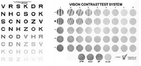
Measurements can be performed with contrast charts. Two commonly used charts are the Pelli-Robson (bottom left) and Vistech (bottom right) contrast charts. Both charts have high contrast letters or gratings on the top left corner. Contrast is gradually reduced towards the bottom right corner – you will be able to discern these if you have high sensitivity.

There is a huge variation as to what is considered ‘normal’ ability to discern contrast. Having one sensitivity measurement in itself is therefore not very useful. What is useful is to perform multiple measurements over a period of time to determine if there has been any change in sensitivity. Testing is currently not routinely performed during childhood vision screening.
The commonest cause for reduced sensitivity to contrast is refractive error (such as myopia and hyperopia). These do not cause permanent visual loss and are usually easily corrected with spectacles or contact lenses.
Contrast testing is quite a specialized test. It is generally not routinely performed in the clinical setting unless there is a specific reason for it. Testing is useful in the context of refractive surgery, particularly in LASIK and photorefractive keratectomy (PRK). Measurements of contrast can be used as another way of assessing visual performance (other than visual acuity) before and after surgery, and also as an aid to help with managing refractive surgery expectations.


