What is endophthalmitis?
Endophthalmitis is inflammation involving the entire eye, meaning that both the front and back portions of the eye are affected. Although the inflammation can be due to various causes, it is generally used to describe an extremely severe infection that has spread throughout the whole eyeball.
This type of infection is devastating. The inflammation causes a great deal of damage to the eye tissues, eventually leading to blindness if not treated appropriately and promptly. It is one of the most serious ophthalmic emergencies and should never be taken lightly. If you suspect that you may have endophthalmitis, please consult your ophthalmologist immediately.

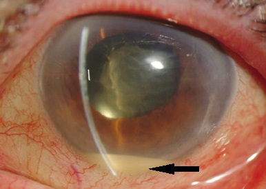
Picture of an eye with endophthalmitis. Note the pus collection in the anterior chamber (hypopyon) associated with a cloudy cornea. This is an indication of very severe inflammation in the eye. If your eye ever looks like this, make sure you contact your ophthalmologist urgently for immediately examination and treatment.
What are the different types?
There are 2 main types of inflammation, depending on the source of the infecting microorganism: endogenous and exogenous.
Endogenous endophthalmitis: This is the infection of the eye by microorganisms that were spread through the bloodstream from a distant infected source, such as from the heart (endocarditis). The typical microorganisms causing endogenous infection are bacteria such as Staphylococcus aureus, Streptococcus pneumoniae, Streptococcus viridans, Escherichia coli and fungi such as Candida.
Endogenous infection is uncommon. It is estimated to happen in 5 out of 10,000 hospitalized patients. It usually occurs in those who are already very ill and are unable to mount an immune response against the infection, such as intravenous drug users, chemotherapy patients and organ transplant receipients taking immunosuppression medications. Other risk factors include AIDS, poorly controlled diabetes mellitus, and long-term steroid use.

The clinical appearance of endogenous endophthalmitis. Inflammation is indicated by the typical yellowish patches on the retina.
Exogenous endophthalmitis: This is the infection of the eye by microorganisms that were directly introduced into the eye either from trauma or from eye surgery. Research has suggested that exogenous infection occurs in up to 11% of open globe eye injuries and approximately 0.1% of eyes undergoing intraocular surgery. Postoperative infective endophthalmitis is one of the most feared complications of eye surgery.
Exogenous infection can be acute or delayed. Acute exogenous infection occurs several days after the event. In contrast, delayed exogenous infection can develop months or even years after surgery, though the average is around 9 months. Delayed infection is less common, but has better prognosis due to the less severe inflammation.
Acute endophthalmitis is exceedingly painful, to the extent that your sleep may be affected. Your vision will drop and your eyelids may swell. Your eye will become very red and inflamed. There will usually be a collection of pus in the front of your eye, which should be easily seen in the mirror.

Acute-onset exogenous endophthalmitis with all the typical clinical features: inflamed conjunctiva, hazy cornea and pus in the anterior chamber (hypopyon; black arrow).
If you have had recent eye surgery or trauma and you experience the symptoms described above, please consult your ophthalmologist as a matter of urgency. How quickly you receive treatment determines how much sight you can save in that eye.
How is endophthalmitis treated?
As the underlying process is that of infection, the immediate priority is to eliminate the infecting organism as quickly as possible. For bacteria, this means antibiotics and for fungi, this means antifungal agents. To identify the infecting microorganism(s), 2 tests are usually performed: vitreous tap and blood culture.
Blood culture: This involves taking a sample of blood from your vein and then sending it for further analysis. Blood culture is particularly useful in cases of endogenous infection.
Vitreous tap: This is a procedure where a small needle is inserted into the vitreous cavity of the eyeball and a sample is withdrawn. The sample is then sent to the labs for culture and analysis to identify the microorganism and the antibiotic or antifungal medication that it is sensitive to. Vitreous tap procedures are performed under local anesthesia and usually combined with injection of antibiotics or antifungals into the vitreous cavity.

When you have been diagnosed with endophthalmitis, you will generally require hospitalization so that treatment can be administered appropriately. You will require multiple eye drops , tablets, intravitreal injections and possibly even intravenous medication.
The eye drops that you will be given are likely to include:
– Antibiotics, such as chloramphenicol or ofloxacin
– Steroids to dampen ocular inflammation, such as dexamethasone
– Cycloplegics to dilate the pupil, such as cyclopentolate
– Lubricant eye drops to protect the ocular surface
Depending on the clinical situation, oral antibiotic and anti-fungal medications may not be appropriate and you may require an intravenous infusion instead. Having infusions into the blood stream requires close monitoring by nursing staff.
You will almost certainly have an intravitreal injection of the antibiotic (such as vancomycin and ceftazidime) or anti-fungal agent (such as amphotericin-B and voriconazole. This is usually performed at the same time as the vitreous tap, but may need to be repeated several times. Injecting the medicines into the vitreous cavity ensures that the drug is delivered directly to where the infecting microorganisms are located.
Sometimes, vitrectomy surgery under general anesthesia is required. This procedure involves removing the infected vitreous gel from the vitreous cavity and replacing the gel with a clear solution. The goal of surgery is to remove as much of the infecting microorganisms as possible. This is done with special instruments, such as the vitrector (or vitreous cutter) and light pipe. Research has found that it is possible to achieve vision of 20/100 or better in up to 74% after vitrectomy.

Endophthalmitis is a devastating condition. If you have had recent eye surgery, make sure you follow the postoperative instructions given by your ophthalmologist in order to minimize the risk of infection. If you are worried in any way, it is best to consult with your ophthalmologist. It is always better to be safe than to be sorry.


